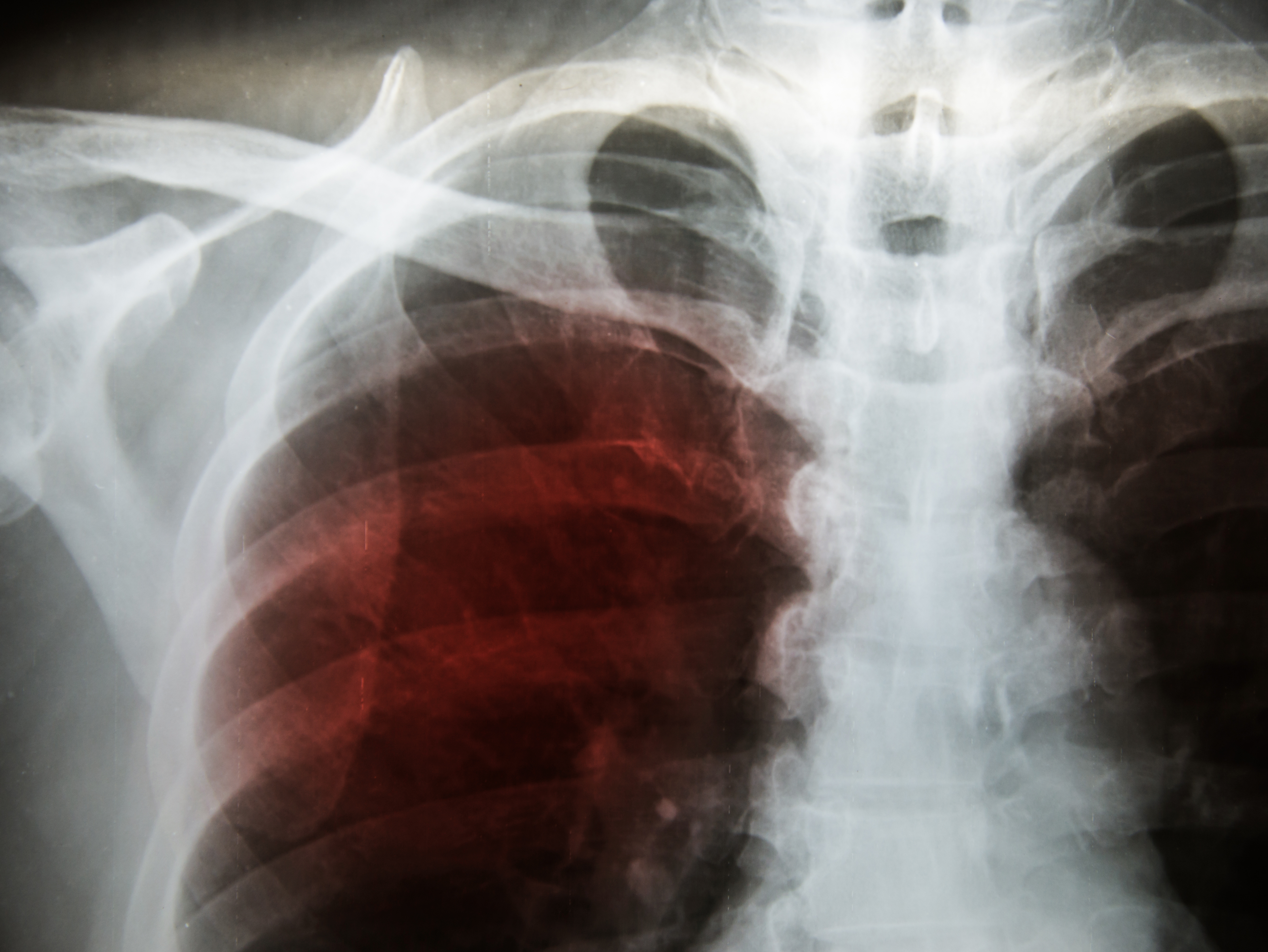
Information on the underlying pathogenesis of fibrotic interstitial lung disease was outlined in a recent report published by Ichimura et al in Proceedings of the National Academy of Sciences. Patients with anti–melanoma differentiation–associated gene 5 (MDA5) antibody–positive dermatomyositis may present with amyopathic dermatomyositis with interstitial lung disease—which can signify rapid disease progression and a high risk of mortality. Using a murine model of interstitial lung disease mediated by autoimmunity against MDA5, the researchers immunized mice four times with recombinant murine MDA5 whole protein alongside complete Freund’s adjuvant once weekly to trigger the production of anti-MDA5 autoantibodies. Although the mice developed MDA5-reactive T cells and anti-MDA5 antibodies, they did not develop full interstitial lung disease. In order to imitate viral infection and acute lung injury, the researchers then administered intranasal polyinosinic-polycytidylic acid in the mice and observed the subsequent development of fibrotic interstitial lung disease, prolonged respiratory inflammation, and fibrotic changes 2 weeks following administration. They found that CD4-positive T cells were critical to the development of interstitial lung disease. Further, upregulation of interleukin-6 mRNA was prolonged in MDA5-immunized mice. However, treatment with anti–CD4-depleting antibodies and anti–interleukin-6 receptor antibodies helped improve the severity of MDA5-induced fibrotic interstitial lung disease, whereas anti–CD8–depleting antibodies were ineffective. The recent findings indicated that autoimmunity against MDA5 may worsen toll-like receptor 3–mediated acute lung injury and prolong inflammation, leading to the development of fibrotic interstitial lung disease. The researchers concluded that interleukin-6 may be an effective therapeutic target for the disease and that further studies may help identify more effective therapies in this patient population.