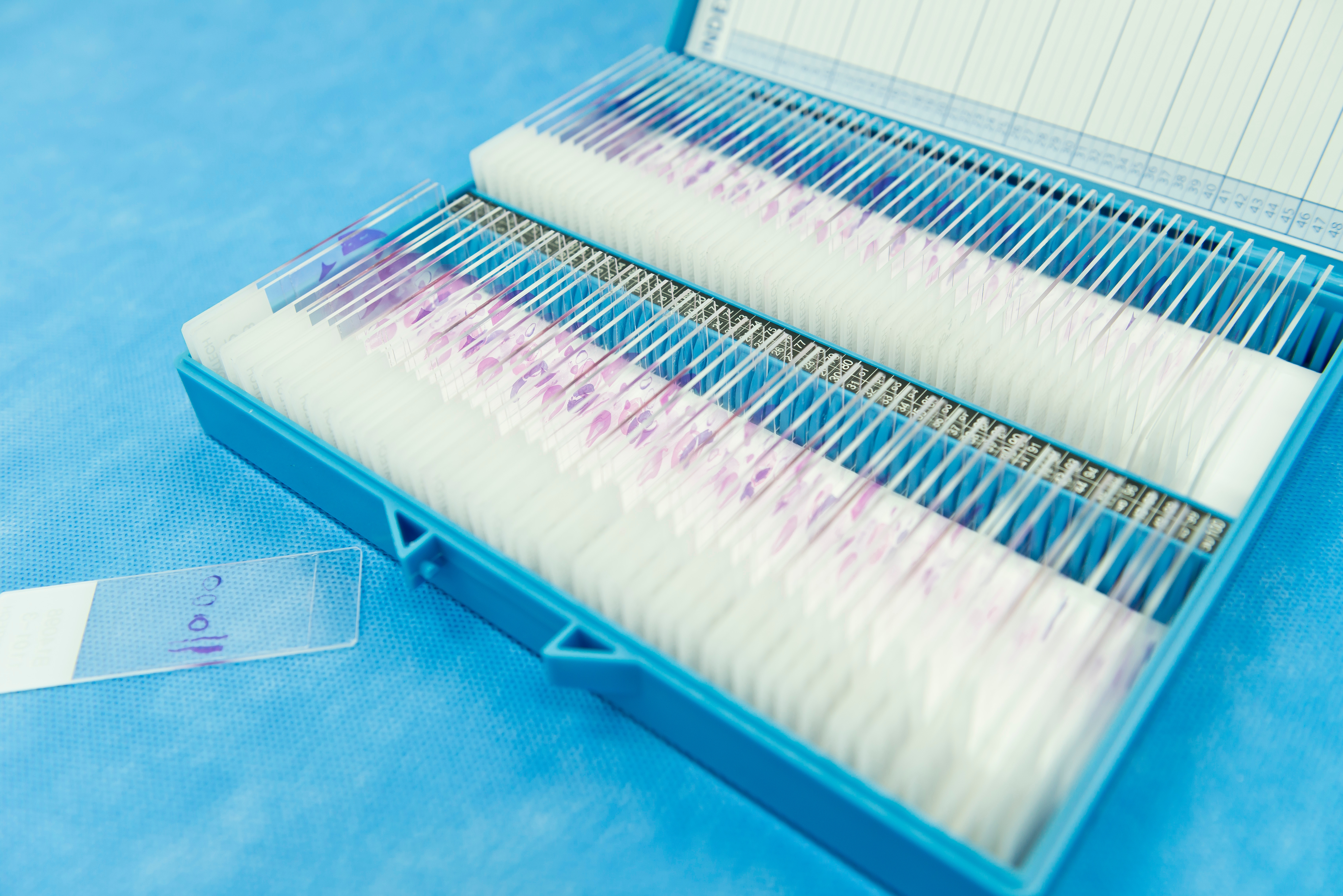
A new method to simplify and enhance skin cancer exams using a novel noninvasive virtual biopsy was described in a recent report published by Winetraub et al in Science Advances. The current standard for diagnosing skin cancer and detecting tumor margins involves the use of histological hematoxylin and eosin–stained tissue sections; however, this strategy is often invasive, labor-intensive, and permanent. In the recent study, researchers developed a micron-accuracy coregistration method known as microregistered optical coherence tomography (OCT) designed to take a two-dimensional hematoxylin and eosin slide and identify the corresponding section in a three-dimensional OCT image using original fresh tissue. The OCT images work by measuring how light waves from a laser interact with tissue to render the insides of the tissue. The researchers then used a paired dataset of over 1,500 hematoxylin and eosin–stained tissue sections and OCT images to train a conditional generative adversarial network to create accurate slides from the OCT data. They demonstrated that the noninvasive OCT images could be converted into high-fidelity two- and three-dimensional virtually stained hematoxylin and eosin slides that imitated those produced by standard biopsy. The researchers reported that they were able to detect the cellular structures on the hematoxylin and eosin–stained tissue sections and OCT images at a similar rate. They emphasized that incorporating the novel OCT imaging strategy into clinical practice may reduce unnecessary biopsies and increase the timeliness of results regarding potentially cancerous skin lesions. In a companion press release on the findings from Stanford Medicine, the study authors concluded: “We’ve not only created something that can replace the current … standard … for diagnosing many conditions, but we … improved the resolution of these scans so much that we started to pick up information that would be [difficult] to see otherwise. This has the potential to transform how we diagnose and monitor concerning skin lesions and diseases in the clinic.”